Muscle Physiology:
Over the next few newsletters, we're going to return to our series on the anatomy and physiology of the human body -- focusing this time on the musculoskeletal system. At first glance, studying muscles and bones might seem boring. After all, who wants to memorize several hundred Latin names? And what do you need to know about muscles and bones to keep them healthy; they pretty much take care of themselves if you eat a good diet, don't they? Eat protein for your muscles and calcium, boron, and vitamin D3 for your bones. There! Done!
In fact, exploring your muscles and bones is far more interesting than it first appears -- and properly taking care of them, far more involved than you might believe. However, if you do things smart and do them right, the rewards can be more than worth the price of admission.
Usually we start by examining the anatomy of an organ or system before we look at its physiology. However, we're going to reverse that order in this case and dive into the physiology of muscle tissue immediately, saving our discussion of anatomy for later. Specifically, we're going to cover:
- The basic functions of muscle tissue.
- The different types of muscle tissue used to perform those functions.
- How muscle is constructed.
- What makes it work.
- How is it powered?
- How can you make it work better?
Basic Facts About Muscles
Because of its high water content, muscle is actually denser than bone. This means, surprisingly, that muscle comprises some 50% of our body weight. It also performs four basic functions. First, muscle provides postural support. That is, our muscles provide stability and postural tone -- to help us stand or hold position, for instance. Muscles also allow us to move or perform work. Third, they contain, position, and regulate the movement of our internal organs -- think peristalsis in our intestinal tract. And finally, muscles are the primary source for generating heat in the body.
Warm blooded animals have to produce heat to survive or they die -- and the colder their environment, the more heat they need to generate. In fact, you have to maintain a narrow range of temperature in your body or there are catastrophic consequences. If your core temperature drops below 93 degrees Fahrenheit, your heart is likely to stop. If your temperature rises above about 108 degrees, proteins in your brain start to denature, causing permanent brain damage.
Effectively, the body loses heat in relation to the square inch surface area of skin it has compared to the cubic volume of muscle mass it possesses. that's why, in general, the smaller the animal, the faster the heart beat -- high ratio of skin surface to low body mass. Hummingbirds, for example, have heart rates when in flight of over 1,100 beats per minute.1 Shrews, the tiniest mammals, have heart rates that can top 1,500 beats a minute.2 Elephants, meanwhile, have heart rates down around 30 beats per minute.3 The reason is very simple. Heat passes out of the body through the skin; thus, the greater the skin surface you have facing the environment, the faster your body loses heat. On the other hand, it's your muscle tissue that is primarily responsible for generating heat in your body. Thus, the greater your muscle mass, the more heat you can generate. (Note: fatty tissue does not generate heat, but it does provide insulation to reduce the rate of heat loss, and it can be broken down and used as fuel by your muscles to generate more heat, but that's a slow inefficient process.) The ability of muscle tissue to generate heat is a very important function of muscles that we normally Don't think about until we're cold and start to shiver. Shivering, by the way, isn't your body's response to the "sensation" of being cold; rather, it's your body's involuntary mechanism for generating heat through rapid muscle contractions. Note: shivering is very different from the shakes we get when we're sick and have fever and chills. Fever shakes are the result of your body getting mixed signals. Your body literally can't figure out if it's hot or cold. The fever tells it that it's hot; the chills, say cold; it's confused. Thus, it shivers in response to the chills even though it's actually already too hot as the result of the fever. (Fever, incidentally, is your body's automatic response to the presence of pathogens as the increased body heat can actually kill some pathogens directly in addition to the fact that heat also stimulates your body's immune system to a heightened response.)
Shivering and shakes are produced in the same way all muscle activity is: by contraction. In fact, all muscle activity is based on contraction. that's all muscle can do -- contract. How do limbs extend, then? The key to more complex actions is that all motion is accomplished by opposing pairs of muscles. Biceps contract to pull your arms up. Triceps, on the other hand, contract to pull your arms down. To clarify our earlier statement, then: all muscle activity is based on opposing contraction. This contraction is initiated by electrical activity. Muscles are highly excitable conductors and respond to electrical stimulation by contracting. In most situations, that electrical stimulation is provided by electrochemical activity in your body. But as anyone who has stuck their finger in a light socket knows, it can also be initiated by outside electrical stimulation.
Types Of Muscle
So far, we've talked about muscles generically and about the things all muscle tissue shares in common. But the fact is that the body actually contains several different types of muscle that despite any outward similarities are distinctly different in their biomechanics and their uses in the body. The three primary types of muscle are:
- Smooth muscle
- Cardiac muscle
- Skeletal muscle
Smooth muscle is a type of non-striated (un-striped) muscle that is found within the tunica media layer of arteries and veins, the bladder, uterus, male and female reproductive tracts, gastrointestinal tract, respiratory tract, and the ciliary muscle and iris muscles of the eye. The glomeruli of the kidneys also contain a smooth muscle-like cell called the mesangial cell. Smooth muscle is fundamentally different from skeletal muscle and cardiac muscle in terms of structure, function, excitation, excitation-coupling, and its mechanism of contraction.
Cardiac muscle, as its name implies, is found in the walls of the heart, where its contractions propel blood both into the heart and then out through the arteries of the circulatory system. It is similar to skeletal muscle in that it is striated. But unlike skeletal muscle, its contractions are primarily involuntary.
Skeletal muscle is a type of striated muscle that is usually attached to the skeleton by tendons. Skeletal muscles are used to hold posture and create movement, by applying force to bones and joints; via contraction. Unlike smooth and cardiac muscle, they generally contract voluntarily (via somatic nerve stimulation), although they can contract involuntarily through reflexes.
Let's now look at each of these muscle types in a little more detail -- devoting most of our attention to skeletal muscles.
Smooth Muscle
Smooth muscle makes up the walls of hollow organs, hair follicles, and blood vessels. It mostly regulates the size of intestinal muscles and glands and plays the primary role in the contractions of the intestinal tract known as peristalsis. Fundamentally, all muscle tissue is built and powered in the same way, with the difference being that smooth muscle is microscopically smooth, not striated. Instead of being grouped in parallel, smooth muscle cells are assembled in irregular bundles of interwoven clusters. It is called smooth because it doesn't have the striations found in skeletal muscle. Surprisingly, considering its assembly from irregular bundles, the final construction of the muscle itself tends to be long and slender. It may be innervated (activated) by one nerve or multiple nerves, depending on function. And it is involuntary.
Cardiac Muscle
Cardiac muscle makes up the walls of the heart. It is microscopically striated, like skeletal muscle, but its striations join together in branching bundles that allow coordinated action. The whole unit contracts together VS the selective contraction found in skeletal muscle. And it is involuntary and auto-rhythmic--although, as yogis have demonstrated, even involuntary muscles such as the heart can be made responsive to voluntary control.
Skeletal Muscle
Although skeletal muscle is similar to cardiac muscle and at first glance looks like heart muscle, it has different characteristics and uses. It is found attached to bone, skin, fascia, and other muscles. Also, any visual similarities to cardiac muscle when viewed with the naked eye disappear when viewed under a microscope. One of the key defining characteristics of skeletal muscle VS cardiac muscle or even the smooth muscle of the intestinal tract is that skeletal muscle is voluntary. That is to say contractions of the skeletal muscle happen when we choose to make them happen -- such as when we lift our arms. Cardiac and smooth muscle, on the other hand as we discussed previously, are primarily involuntary. For most people, their hearts tend to beat--or not--no matter what they think about it.
Skeletal muscle looks like it is made up of a series of stripes, which is why it is also sometimes called striated muscle. It has this appearance because it is comprised of a series of long bundled threads of muscle known as myofibrils. It is this bundling together of several groups of fibers in parallel that gives skeletal muscle its striated appearance. Incidentally, myo is Greek for "muscle."
A single muscle cell is a compound structure composed of several bundles of myofibrils that contain myofilaments. The myofibrils have distinct, repeating microanatomical units, known as sarcomeres, which represent the basic contractile units of the myocyte or muscle cell. (The word sarcomere comes from the Greek: sarco for "flesh" and mere for "part" -- thus "flesh part.")
The appearance of striation is further reinforced by the fact that all of these individual muscle cells and fibers are grouped into parallel bundles known as fascicles, which are wrapped up in a smooth, slippery covering of connective tissue, known as the perimysium. Those bundles are then grouped together and wrapped in a smooth envelope of connective tissue known as the epimysium to form what we know as a single muscle -- such as a tricep. From smallest to largest, the parts of a muscle are as follows:
- Sarcomere (the basic contracting unit of the myofilament).
- Myofilament.
- Myofibril.
- Myocyte (a single muscle cell), also referred to as a "muscle fiber."
- Fascicle (a bundle of 10-100 muscle fibers).
- Perimysium (the layer of connective tissue that surrounds a fascicle).
- A single muscle -- such as a bicep.
- Epimysium: The outermost layer of fascia, consisting largely of collagen, surrounding a whole muscle and keeping it distinct from--and allowing it to slide over--adjacent muscle groups.
Muscle contraction happens at the level of the sarcomere. The sarcomere is comprised of two protein filaments -- the thick filament is made of the protein myosin and the thin filament made of the protein actin. (Incidentally, that's why protein consumption is important in muscle development. Protein is the fundamental building block in muscle.) When triggered by nerve impulses and powered by ATP, the thick myosin filaments contract. This contraction is transmitted to the thin actin filaments by a series of microscopic spurs in both the thick and thin filaments that ratchet into each other. The thin filaments then transmit the force generated by myosin to the ends of sarcomeres where they are attached and then all along the length of the myofilaments to contract the entire muscle fiber. This is the fundamental element of muscle contraction. Just multiply this out over countless sarcomeres, repeated through vast numbers of fibrils, and numerous muscle fibers, and you have muscle movement. (In a little bit, we'll talk about how this contraction is powered.)
Motor Unit
Specific nerves stimulate a specific group of fibers (or muscle cells) referred to as a motor unit. Essentially, a motor unit is composed of a motor neuron and the muscle fibers (cells) it activates. The innervation (stimulation) by a motor neuron may activate as few as 10 muscle fibers to as many as 2,000. The size of that group is determined by the needs of the muscle group in question. The groups are very small in the muscles of the eyes and fingers, for example. This allows for very fine control of those muscles. Groupings in the muscles of the back and thighs, on the other hand, are much larger since there is little need for fine control in those muscles. The signal to activate travels down from the brain, through the spinal cord, and then out to the group of fibers in question. The activating neuron connects to the muscle bundle at what is known as the neuromuscular junction (NMJ). Because most neuromuscular junctions are located in the middle of the muscle fiber, the wave spreads from the middle outward toward the end of the fiber, allowing the muscle to make a smooth contraction. The nerve cell body is located in the spinal cord, with the filament like axon traveling out from the spinal cord to the muscle fibers it controls. The brain can selectively fire muscle fibers as needed by choosing the motor neuron units it wants to fire. All fibers in a motor unit fire if a signal is transmitted through the motor neuron to that unit. Note: the axons are surrounded by a fatty, insulating coating known as the myelin sheath that facilitates the transmission of nerve impulses along the axon and that prevents any leakage of that signal or extraneous signals from disrupting the transmission. Keeping the myelin sheath intact is essential for proper muscle function.
The NMJ actually consists of a microscopic empty space known as a synaptic gap. The electrical impulse of the nerve cannot cross this gap on its own. In place of direct electrical stimulation of the muscle by the nerves, the impulse is carried across the gap by a chemical intermediary: the neurotransmitter. The primary neurotransmitter used in the stimulation of muscle motor units is acetylcholine (Ach), which is produced in synaptic vesicles located in the bulb at the end of the axon. The motor end plate is the region on the muscle side of the synaptic gap in proximity to the axon terminal bulb. The motor end plate contains the Ach receptors, which when touched by the Ach molecule, fire the muscle to contract. Sufficient supplies of acetylcholine are necessary in the synaptic gap for proper muscle function. Ach is carried across the gap by the sodium pump mechanism that we explored in detail when examining the intestinal tract. Thus, maintaining proper hydration and electrolyte balance is essential for proper muscle function.
Acetylcholinesterase is the enzyme that breaks down the Ach and ends the contraction stimulus so the muscle doesn't spasm. After the process is stopped, there is a refractory period--a period of recovery--in which the muscle is pumping out the sodium, reestablishing the sodium/potassium balance, and repolarizing the charges on either side of the gap--during which no further stimulation is allowed before the muscle fiber can fire again. Fortunately, we have millions of fibers and they're not all firing together so that while some fibers are recovering, others are ready to go.
Powering Muscles: The ADP-ATP Energy Cycle
Energy is stored in the muscle cells in the form of adenosine triphosphate (ATP). The energy is released when the last phosphate group is split off, converting the ATP into adenosine diphosphate (ADP). The released energy is used to power muscles and do work. This ATP-ADP energy cycle is what powers the ratcheting action in the myosin bands in each individual sarcomere. ADP then needs to be converted back to ATP for another round of work. Rebuilding ATP from ADP requires energy to be pumped back into the system, which is primarily provided by glucose through a process known as glycolysis. Glycolysis is up to 15 times more efficient if oxygen is present.4 Anaerobic glycolysis (without oxygen) will produce 2 ATP molecules for every molecule of glucose. But aerobic glycolysis produces 29-30 molecules of ATP for every molecule of glucose. This means that maintaining sufficient oxygenation of muscle tissue is essential for maximum muscle efficiency.
Note: ATP is made up of an adenosine molecule with three attached phosphate groups. Adenosine itself is comprised of a molecule of adenine (one of the key components of your DNA) attached to a ribose sugar. This makes having a sufficient supply of ribose available important for maximum muscle efficiency.
Conclusion
Let's conclude by detailing those supplements you might want to consider to improve both the capacity of your body to build muscle and its ability to support and maximize the effectiveness of those muscles it already has.
To Build Muscle Tissue:
- HGH secretagogues are precursors necessary for the production of human growth hormone in the body. Typically, such formulas contain ingredients such as glutamine, tyrosine, GABA, arginine, and lysine. HGH promotes the growth of lean muscle in the body, and levels of HGH drop as you age. You Don't want HGH injections for a number of reasons, but a secretagogue supplement will help increase HGH production -- as will exercise.
- Protein. Muscle is built from protein, so obviously you need protein to build muscle. But you Don't need massive amounts unless you're doing extreme levels of exercise, and it doesn't have to be meat or dairy protein.
- Testosterone unbinders. Testosterone is the hormone that tells your body to use calories to build muscle and not store them as fat. On the other hand, you Don't really want to take additional testosterone, since higher levels of testosterone are associated with a shorter life expectancy in some studies.5 Then again, other studies show that low levels of testosterone shorten lifespan.6 (let's hear it for contradictory studies.) Instead, what you want to do is free up the testosterone you already have so that it works more effectively for you. That gives you all of the reward without the risk.
- Carnosine based formulas to protect that muscle from the destructive effects of glycation. Supplementation with carnosine has the additional benefit of dramtically speeding up recovery time after intense workouts.
- Arginine works by filling your muscles with water and nutrients. When taken consistently, it helps your body maintain lean muscle mass and promotes the release of growth hormones.
To Allow Contiguous Muscle Groups To Easily Slide Over Each Other A Healthy Epimysium:
- Sufficient hydration. When determining how much water you need, the medical community rarely looks deeply enough and therefore comes to the wrong conclusions. Early stages of dehydration show up in a "sticky" epimysium, which means that muscles no longer glide smoothly over each other.
- Collagen. The epimysium is made largely of collagen. Supplementation with oral collagen supplements helps keep the epimysium intact.
- ASU. Avocado soy unsaponifiables stimulate the production of collagen in the body.
- Systemic proteolytic enzymes help break down any protein adhesions that may form and cause muscles to "stick" together.
- Deep muscle bodywork such as BioSync helps keep the muscle planes from sticking together -- and helps separate them if they're already stuck.
- Yoga. The total body stretches offered in yoga work every muscle plane against every other plane--helping to keep muscle planes moving freely and prevent any adhesions from forming between planes.
To Power The Nerve Impulses That Drive Muscle Activity:
- Acetylcholine. DMAE is a precursor for the production of acetylcholine in the body.
- The myelin sheath is comprised of 70 percent fats and cholesterol and 30 percent protein. It is easily damaged by:
- An accumulation of toxic heavy metals. Detox heavy metals regularly.
- Inflammation in surrounding tissue, which can damage the myelin sheath. Reduce systemic inflammation using proteolytic enzymes
- A deficiency of methylation nutrients compromises the ability of the myelin sheath to repair itself.7 Supplementation with any and all of the methylation nutrients such as folic acid, B-12, TMG (tri-methyl-glycine), and SAMe can be helpful.8
- Vitamin D protects against demyelination.9
- Omega-3 fatty acids
Power The ATP-ADP Energy Process
- CoQ10 and CoQ1 (NADH) are the key members of the electron transfer chain in mitochondria (the primary engines of almost all bioenergy production). The passage of electrons along the electron transport chain is coupled to the formation of ATP. Coenzyme Q-10 needs NADH to be effective.
- Your body generates ATP from D-Ribose, which is normally made from glucose. However if the cell is lacking in energy, then the cell converts the glucose to lactic acid instead of D-Ribose. D-ribose as a nutritional supplement is useful because it is immediately available for the generation of new ATP. Also, sufficient supplies of D-ribose minimize the production of lactic acid.
- Nicotinamide (vitamin B3) plays an important role in the synthesis of components necessary for the production of ATP.
- PQQ (Pyrroloquinoline quinone) is taken as a dietary supplement to support mitochondrial health and cellular energy production and to protect the body from oxidative stress. Even better, it can stimulate production of new mitochondria -- where ATP is produced.10
- Creatine is found naturally in vertebrates and helps to supply energy to all cells in the body.
- Proteolytic enzymes are beneficial here too. Since they improve the blood's ability to take up oxygen and carry out carbon dioxide, they increase oxygen levels in cells -- thus dramatically improving the production of ATP through glycolysis.
That's it for now. In our next newsletter, we'll explore the anatomy of the muscular system and how different types of exercise promote the growth of different types of skeletal muscle.
Written By: Jon Barron
Website: www.jonbarron.org
Dated 1/28/2013
Human Anatomy & Physics Of Muscles
In our last newsletter, we examined the physiology of the human muscle system from an alternative health perspective. In this newsletter, we continue with that theme by exploring both the physics of muscle movement and the code that underpins muscle anatomy. And no, the goal is not to teach you the names of all 799 skeletal muscles in the body, but rather to show you "how" the muscles are named. Muscle names are not just a bunch of random Latin words, but rather, are named according to a set of informal rules; and once you understand those rules, you can pretty much tell where any muscle is located and what it does.
By itself, though, this is not important; but once armed with the information, we will then be able to talk about exercise -- and exactly how to exercise our muscles to accomplish specific goals and achieve optimum health.
The Physics Of Muscles
Before we can discuss the naming system for muscles, we need to understand the principle of levers that make the whole musculoskeletal system work since many muscle names are derived from that principle. The reason for this is simple: all skeletal muscles provide stability and produce movement in the body by acting as the force or effort applied to the levers of our bones -- and by using opposing forces to achieve mechanical advantage. Or to put it another way, muscles move bones around joints.
There are actually three types or classes of levers, but when we think of levers, we normally think of what are known as first class levers. These are the levers where the fulcrum is located between the effort (or force) we exert and the load (or weight) we are trying to move.
With a lever, you can either increase the force of the movement, its speed, or its range of motion -- but you can't do all three at the same time. Levers are about tradeoffs; you trade one benefit for another. For example, in the above lever, if we move the fulcrum to the right, we gain speed and range of motion since it takes very little movement on our part to move the load rapidly through a great distance. However, in exchange for this advantage, it does take a great deal of effort.
On the other hand, if we move the fulcrum to the left, closer to the load, it increases our strength significantly, but we get much less movement out of our load.
To clarify, if the distance from where we exert effort to the fulcrum is 10X's the distance from the fulcrum to the weight, we can lift 10 lbs with 1 lb of force. Or vice versa. This is what Archimedes was referring to when he said, "Give me a lever long enough and a place to stand, and I can move the world."
In truth, there are very few first class levers in the human body -- the muscles of the back of the neck being a notable exception. As you can see, the muscle is attached to the clavicle (collarbone) and the back of the head. The fulcrum lies at the atlas bone at the top of the spine. The load is the front of the head that we're trying to hold up. There's very little mechanical advantage here since the fulcrum is pretty much centered between the load (the front of the head) and where the muscle exerts its force (the back of the head)--but then there doesn't really have to be much gain or loss since these are essentially postural muscles that merely hold the head up. You know exactly which muscles these are if you've ever tried to sleep on a plane and your head keeps dropping down and walking you up when the muscles relax.
Second Class Lever
In a second class lever, the load is located between the fulcrum and where the effort is applied. This is like a wheelbarrow.
There are very few second class levers in the body. The muscles used when you stand on your tiptoes would be an exception. Your toes are the fulcrum; your body is the load; and your calf muscles pulling on your heel provide the effort. There is very little mechanical advantage when standing on our toes because the resistance is somewhat removed from the fulcrum, but we get to lift a pretty good distance with minimal muscle movement.
Third Class Lever
Well, there are only three classes of levers, and if the human body has very few first and second class levers, then by the process of elimination, most of the muscles in our body must work as part of third class lever systems. And that is exactly the case. In a third class lever system, the force is applied between the fulcrum and the load.
While you might think that all muscles would be designed to increase strength--ala Archimedes-- that's not necessarily the case. Some of the major third class levers in our body are designed to increase range of movement and speed at the expense of strength. Think of your biceps for a moment -- think of how little muscle movement is required to fully bend your arm. that's a lot of motion, for very little muscle movement. When doing curls for example, the elbow is the fulcrum, the weight in your hand is the load, and biceps muscle is the force. Again, the gain is in speed and range of motion. that's why it takes so much work at the gym to build strong biceps. Your thighs are similar. Speed and range are gained over strength -- which is why the thigh muscles need to be so large to compensate.
Basic Terminology In Understanding Muscle Anatomy
Before we actually get to the naming of muscles, we need to touch on a few pieces of terminology that are important to know.
Proximal means that something is near the top of an organ or muscle. (For example, when talking about the biceps, up near where the bicep attaches to the shoulder is proximal.)
Distal means that something is away from the top of the organ or muscle. (Down, just below the elbow where the bicep attaches to the forearm is distal.)
The origin of a muscle is where the muscle attaches to the more stationary bone or structure. (Again, using the bicep as a reference, the origin is at the shoulder, the more stationary point of attachment.)
Its counterpart is the insertion, which is where the tendon at the end of the muscle attaches to the moving bone or structure. (For the bicep, insertion is on the radius of the forearm, near the elbow, the part of the arm that we're trying to lift up as the bicep contracts.)
Tendons are the dense connective tissue (thickened, fibrous structure made of collagen) at the ends of every muscle group that connect the muscle to bone. (Note: Ligaments are not involved with muscle. They attach bone to bone to stabilize joints. For example, think of the ligaments stabilizing the knee -- and who doesn't know about those ligaments thanks to American football.)
The tubercle is the thickened area of a bone where a tendon attaches. It is thickened because bone growth has responded to the increased stress at the area of attachment. (Note: any stress on a bone causes the stressed area to thicken and grow stronger. This is an important concept to remember when it comes to exercise and maximizing bone strength.)
And the belly of a muscle is the thicker middle portion of a muscle. (The part that bodybuilders are trying to make look really big--as in the bicep.)
The Eight Part Code For The Naming Of Muscles
Now let's talk about how muscles are named. Surprisingly, virtually all muscles are named based on one or more of the following eight criteria. Learn these eight criteria--and their Latin (or sometimes Greek and even French) equivalents--and you can locate in the body virtually any muscle that someone names. And thus, unless you're an anatomist, you Don't have to memorize all 799 muscles (approximately) to be anatomically conversant. The eight criteria are:
- Size
- Shape
- Orientation of the muscle fibers
- Mechanical action of the muscles
- Number of origins of a muscle (and yes, muscles can have more than one origin)
- The points of origin and insertion
- Name of the muscle function
- The muscle's location
Size
When it comes to size, muscles are large or small, short or long, or wide.
The largest muscle in a related group of muscles is often referred to as maximus or magnus. An example that you're familiar with is the gluteus maximus. Gluteus is Latin for your rear end -- or more politely, your buttock. Thus, gluteus maximus identifies the largest muscle in your butt--the one you feel when you squeeze the cheeks of your butt together. Another example is the adductor magnus, which is the large muscle running down the inner thigh that pulls the leg back in from the side. You can feel this muscle if you balance against a table, putting your hand against the inside of the opposite thigh and then resisting as that hand tries to push the leg out to the side.
Minimus, not surprisingly, refers to the smallest of a group. Thus gluteus minimus identifies the smaller butt muscle located underneath its maximus big brother.
Longus, as you might suspect, refers to the longest of a group--as in the adductor longus, which is thinner than the adductor magnus and runs essentially parallel to it.
Brevis identifies the shortest of a group. The adductor brevis runs across the thigh to assist in pulling the thigh in towards your body's midline as opposed to down the length of your inner thigh as do the adductor magnus and minimus.
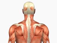
In Latin, the word "latus" means "side." Thus latissimus identifies the largest muscle "in width" in a group. Latissimus dorsi is the name of the large muscles that run from under your arms, across your "sides," and then across the middle of your back. Bodybuilders refer to these as their "lats."
So again, quick review, when it comes to size, the key identifiers are maximus or magnus, minimus, longus, brevis, and latissimus.
Shape
There are really only four shapes that you need to concern yourself with when it comes to naming muscles: trapezoids, triangles, saw tooth, and flat.
A trapezoid is like a rectangle, but with only two sides that are parallel as opposed to all four. For simplicity you can think of it as an oddly shaped rectangle. The trapezius is the large distorted rectangle that sits on the upper portion of the back and is rotated 45% so that one corner attaches to each shoulder, one at the top of the neck, and the fourth corner to the middle of the back. I actually think it looks more diamond shaped, but I didn't discover it, so I didn't get to name it.
The fourth letter in the Greek alphabet is delta, which is drawn in the shape of a triangle. Deltoid, then, refers to the triangular shaped muscle that sits on the top of the shoulder.
The Latin verb for saw is "serrare." In anatomy, then, serratus means saw-toothed in shape. Serratus anterior is the name of the muscle that holds your scapula (shoulder blade) to your ribs. It gets its saw-like appearance from the pattern it makes is it attaches to each individual rib.
The French word for flat is "plat." Think of the word plateau. The platysma muscle is the broad, thin, flat muscle that has its origin across the middle of your chest, runs up across the front and sides of your neck and ends at your chin. Its primary role is to hold the other muscles of the neck in place and help with facial expressions. It comes into play when you tense your jaw and neck.
Orientation Of The Muscle Fibers
Orientation refers to how the muscle fibers line up to the midline of the body. This makes it pretty simple. They can only be parallel, perpendicular, or diagonal to that line.
Rectus is the word used to identify those muscles whose fibers run parallel to the midline. The Latin word for "right" is "rectus." Rectus abdominis, for example, identifies the six-pack abdominal muscle that runs down the front of the stomach--with its fibers running parallel to the spine. I have to admit, getting parallel out of rectus is a bit of a stretch. Its original root, as I mentioned, is in the Latin word for "right or true." Think of the word rectitude which identifies the quality of being virtuous or straight. Well, from that you get "rectus" as identifying a muscle that runs straight and true -- or dare I say: parallel. Don't blame me. Nobody ever said all doctors were Latin scholars. These are the famous "six-pack" muscles. They get that name because, when well developed, the muscle gets segmented by bands of cartilage that give the muscle the appearance of having six separate sections.
Oblique comes from the old French and means "at an angle." Thus we have both the external and internal obliques of the abdomen, which run one on top of the other, with their fibers running at an angle to the midline and perpendicular to each other. These are the muscles just to the outside of your six-pack, and these are the muscles you work when you do sit ups and touch each elbow to the opposite leg.
Transverse means crosswise or perpendicular to the midline of the body. Thus we have the transversus abdominis. From the name, you can pretty much guess that they're located in the abdomen and which direction their fibers run. In fact, these muscles are located in the sides of the abdomen, underneath the obliques. And the fibers do indeed run across the body. And it's these muscles you feel when you stretch from side to side.
Mechanical Action Of The Muscle
Here we're talking about what the muscle does. Does it flex, extend, turn up, turn something down, lift it up, lower it, rotate around a joint, move body parts away from a midline or pull them back towards the midline, make an area rigid, or close an opening? The action that a muscle performs is often used in naming that muscle.
Flexor muscles decrease the angle at a joint. The flexor pollicis longus is the muscle in the forearm that pulls on the thumb and bends or flexes it inward toward the palm. We already know that longus means it's the longest muscle in its group -- and, in fact, it runs the full length of the forearm from the elbow to the thumb. And "pollicis" is Latin for thumb. Thus, the name tells us that it's the long muscle that flexes the thumb.
Extensors are the muscles that counter flexors. They increase the angle at a joint. The extensor pollicis longus, therefore, is the long muscle in the forearm that straightens out the thumb once it's been bent inward.
Pronators turn limbs so that they face downwards or backwards. If you hold your arm out in front of you, palm up, it's the pronator teres muscle that allows you to turn the arm at the elbow so that the palm is now facing downwards. The Latin word "pronus" means "face down" -- as in lying prone. And "teres" is Latin for "rounded or cylindrical," which refers to the shape of the muscle.
The counter to a pronator is a supinator. The musculus supinator, for example, is the muscle in your forearm that turns your palm back facing up after you've pronated it down. Supinator comes from the Latin word supinum, which means "lying on your back."
Levators, as is obvious from the word, are muscles that lift things up. The levator ani is the muscle that pulls the anus up at the end of the digestive tract. If that muscle starts to weaken, you can feel the anus starting to push out and down -- very uncomfortable.
Depressors, as would be expected, are the opposite of levators; they push or pull things downward. The depressor labii inferioris is the muscle located below (or inferior) to the lips that pulls the lower lip down.
Rotators, as the name implies, produce a circular movement around a joint -- the rotator cuff in the shoulder being the most obvious example.
Abductor muscles move bones away from a midline in the body. The term is used both generically to describe the action of any muscle that moves away from the midline (the gluteus medius, for example, is an abductor in that its action is to pull the thigh out from the midline) and as part of the formal name of a handful of muscles such as the abductor pollicis brevis, which pulls the thumb away from the palm.
Adductors move the bones back towards the midline as we saw with both the adductor longus and the adductor brevis, which are located in the inner thigh and that we looked at previously.
Tensor muscles make things rigid. The tensor fascia lata muscle in the leg tightens and gets rigid to support the knee.
Sphincters close openings, as does the anal sphincter.
Number Of Points Of Origin
A small number of muscles are named after the number of points of attachment they have at their points of origin. These would be:
The biceps brachii in your arm that has two points of origin. Brachii is Latin for branches -- thus, your biceps muscle has two branches at its origin.
The triceps brachii in your arm has three points of origin.
And the quadriceps in your leg has four points of origin.
Origin And Insertion
An even smaller number of muscles are named after the parts of the body where they start and end -- their origin and insertion. The sternocleidomastoid muscle, for example, originates from both the sternum and clavicle (breastbone and collarbone) and inserts into the mastoid bone (just below the ear).
Named By Function
And a small number of muscles are named after their function.
The risorius is a facial muscle that is crucial for the expressions of smiling and laughter. Humans are the only animals that have a well developed risorius. Risorius comes from the Latin word "risus," which means "laugh."
Masseter comes from the Greek word for "chew," and that's its function in the human jaw.
The sartorius muscle (from the Latin word for "tailor") runs from the outer hip, across the thigh, and ends at the inner knee. This muscle pulls the leg up at the knee while simultaneously turning it inward. It is used to cross the legs in the manner of an old time tailor sitting on the floor and sewing hides together--hence the name.
Location
And finally, some muscles are named by where they are found in the body.
The temporalis muscle is named after the temporal bone (your temple) on top of which it is located.
Likewise, the zygomatic bone is your cheekbone, and the zygomaticus muscles are located over your cheekbone.
Review Of The Major Muscle Groups
Now that we know how muscles are named, let's quickly work through the body and take a look at the major muscle groups
Upper Body Front
Pectoralis major muscles originate in the sternum and clavicle. They attach under the arm and pull the arms up across the body and rotate them inward. These are your pecs. They are worked by butterflies and bench presses.
The deltoid originates in the clavicle, with insertion in the arm below the shoulder. The deltoid elevates the arm when it contracts.
Biceps brachii and brachialis work in tandem, attaching in the forearm, and lifting the forearm.
Rectus abdominis is a paired muscle that runs down either side of the abdomen. It is supported by tendons that give the muscle the appearance of being segmented -- thus the six pack.
Thigh Muscles
- Sartorius, as discussed previously
- Adductor magnus and longus, which pull the legs together at the midline
- Rectus femoris, which helps you kick
- Vastus lateralis and vastus medialis, which along with the rectus femoris help extend the knee and form a cage around it.
Back
The trapezius and latissimus dorsi, joined by a mass of tendons, pull across the back.
Under the scapula are the Infraspinatus, teres minor, and teres major, which all serve to stabilize the shoulder blades so that the shoulders can move.
Gluteus maximus and the gluteus minimus hidden underneath the maximus are primarily stabilizing muscles. They support the pelvis and the trunk where it sits on the femur (the thigh bone). These muscles are most noticeable when standing on one leg. Without special exercise--squats, lunges, and backward leg lifts--they tend to lose tone as we age. This is the saggy butt often associated with aging.
Hamstrings: semitendinosus and biceps femoris (not to be confused with the biceps brachii in your arm) -- to flex the lower leg at the knee. (Semitendinosius comes from the Latin words for "half tightly stretched" and refers to normal tightness of the calf muscles.)
Calf muscles: gastrocnemius, with its two heads -- medial and lateral. (Gastrocnemius comes from the Greek and Latin words for calf.)
Face And Neck
- Frontalis wrinkles your brow.
- Orbicularis oculi circles the eye.
- Muscles of facial expression:
- Levator labii superioris, which raises the lips from above.
- Zygomaticus minor and major that raises the lips from the side.
- Depressor anguli oris that pulls the lips down.
- Risoris that makes you smile.
- Mentalis that pulls the lip down with the chin.
- Neck muscles:
- Sternocleidomastoid muscles flex and rotate the head.
- Omohyoid muscles are used primarily in chewing and swallowing.
- Sternohyoid helps control the hyoid bone, which plays a key role in swallowing and speech.
- Thyroid cartilage.
Conclusion
let's look back at a couple of key pieces of information that we explored today and see what principles we can draw from them that might be useful in optimizing our own health.
Developing Muscles In Balance
First, early in the newsletter we explored in some detail that all muscle movement is based on the principle of levers, activated by contracting muscles -- and that to get movement in opposite directions, we need opposing pairs of muscles that pull the levers in opposite directions when they contract. Thus, biceps bend our arms up and triceps straighten them out. This brings us to our first key concept: balance.
When you work one set of muscles--say the biceps--you need to work their counterparts--in this case, the triceps. This is not just a matter of looking pretty for bodybuilding competitions; it's a question of long term fitness. When muscles are not developed in balance:
It stresses bones in only one direction, which causes bones to build in an unsound manner. Remember, bones build in response to stress--both gravitational and exercise induced. All muscles attach to bones and pull on those bones, using those bones as levers. If, for example, you continually work your biceps without developing your triceps, your muscles are overwhelming pulling on your arm bones in only one direction. This causes the bone to develop strength on one side, and weakness on the other. (we'll soon explore how bones build in response to stress, which is why weight bearing exercise is so important.) Prolonged, uneven stress on bone is a prescription for serious bone curvature or even a predisposition for breakage down the road.
It also should be noted that developing muscles that are out of balance is a prescription for chronic muscle aches and pains as the over developed muscles continually pull and tweak their underdeveloped counterparts.
And it's bad for spinal alignment. If your core muscles are not developed equally (or not at all), they will pull the spine in one direction or the other, rather than stabilize it in the proper alignment. This guarantees chronic back pain and even disc problems in time.
Muscle Cages
Our muscles also tend to work as protective cages in several areas of our body. These cages not only protect those sensitive areas from damage, they also support and stabilize those areas to protect them from going out of alignment and chronic pain. Two key areas stand out: your lower back and your knee.
The core muscles of your stomach (rectus abdominis, transversus abdominis, and the obliques) and your back (the latissimus dorsi) not only protect your internal organs and hold them in place; they also literally hold your spine in correct alignment. If those muscles weaken or develop an imbalance, you can pretty much guarantee, you'll be dealing with chronic back pain.
As for your knee, a cage of muscles and their tendons runs from the thigh to the lower leg protecting the knee from instability and injury. Damage to these muscles and tendons requires extensive physical therapy; otherwise the knee will never recover its full strength. Medial and lateral ligaments also help stabilize the knee, as do anterior and posterior ligaments -- all of which can be injured. The real protection for your knee, though, is this cage of tendons that come from the muscles of your thigh. The stronger those muscles are the stronger that cage is. And keeping in mind that painful knees are one of the biggest reasons people have difficulty walking as they age, keeping those muscles toned and in balance makes all the sense in the world. And this is especially important after any injury--no matter your age. For example, when you are injured and become immobile, the huge muscles of the thigh, such as the quadriceps are the first muscles to atrophy. Without extensive physical therapy, they will never fully recover, which means that your knee is now more prone to injury because its cage is compromised.
Next Newsletter
In the next issue, we'll conclude our examination of human musculature by talking about the principles of exercise. You will want to read this newsletter if you are concerned about your health. Muscles are not just for good looks. They:
- Give us strength.
- Make bones stronger.
- Hold your spine in line and your organs in place.
- Protect you from falling. The stronger your legs muscles are, the less likely you are to be unsteady and fall as you age.
- Protect you even if you do fall. Muscle is dense tissue. If your hip is surrounded by toned muscle, when you fall, it cushions your hip bone and protects it from breaking.
- Keep you independent longer as you age.
Specifically, we'll explore the different types of muscle fibers we have and how certain exercises can selectively change the balance of those fibers in our muscles -- emphasizing one kind of fiber over another. it's what allows one set of exercise to develop a group of muscles that are suitable for a bodybuilder, while another set makes the same group of muscles suitable for a sprinter. And for those of us who are not professional athletes, understanding muscle fibers and how to exercise them is what allows us to tune our bodies to maximize the lifestyle that we actually lead -- assuming we're not couch potatoes.


